![]()
Cardiology and techniques to detect cardiac diseases
in rabbits
Esther van Praag Ph.D.
|
MediRabbit.com is funded solely by the
generosity of donors.
Every donation, no matter what
the size, is appreciated and will aid in the continuing research of medical care
and health of rabbits.
Thank you
|

Warning: this page contains pictures that may be distressing for some persons.
Cardiology in pet rabbits is a
domain on which little information is available. Therefore, the incidence of
cardiac problems is not well known.
Although information is scarce,
it is possible to diagnose the problem by means of a complete cardiac study
including radiography, electrocardiography and/or ultrasound analysis to
treat it the cardiac disorder appropriately.
The
heart is located in the thoracic cavity with the apex (tip of the heart)
directed backward and slightly to the left; the base is directed forwards. As
observed in other small animals, the rabbit heart has 4 chambers: 2 auricles
and 2 ventricles separated by inter-auricular and inter-ventricular septa. It
has, furthermore, some anatomical particularities:
• Right and
left ventricles that form the muscular caudal portion of the rabbit heart.
The left ventricle is larger than the right one; they are separated from each
other by the interventricular septum. Their walls are raised into muscular
ridges. The right ventricle is much thicker than the right auricle and forms
the right side of the conical apical portion, but without reaching the apex.
It gives off the pulmonary artery in front. Cusp valves separate the
ventricular chambers from the pulmonary artery and the aorta.
The
ventricular chambers are separated from the auricles by flap valves, which
are held in place by tendons. The heart valve between the right auricle and
right ventricle (tricuspid valve) possesses only two cusps, and not three as
usually observed in other animals.
• Right and
left auricles, which are located in the cranial part of the heart. They are
small chambers that receive the venous blood from:
• The
cranial and caudal vena cava (one of two large veins returning blood from
outer parts of the body to the right chamber of the heart) and the coronary
sinus (receiving blood from the heart itself), which are drained into the
right auricle,
• The left
and right pulmonary veins, that bring oxygenated blood from the lungs, open
together into the cavity, on the dorsal side of the left auricle. Each auricle
possesses, in addition, small muscular flaps.
The
sino-atrial node - or pacemaker from which originates the heartbeat - is
located in the wall of the right auricle.
Further
physiological points differentiate the rabbit heart from that of other small
animals:
• The
aortic nerve has no chemoreceptors, but only baroreceptors. This means that
it does not have sensory nerve cells that are activated by chemicals, but
only pressure-sensitive nerve endings, that stimulate reflex mechanisms that
allow the body to adapt to changes in blood pressure by dilating or
constricting the blood vessels.
• The
pulmonary artery and its branches are heavily muscular.
• The
coronary arteries, which supply the cardiac muscle and are given off from the
aorta, can easily be compressed, leading to ischemia of the myocardium, due
to poor collateral circulation.
Rabbit cardiac
parameters
Rabbit abdominal
radiography
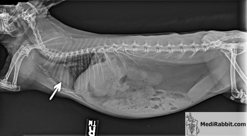
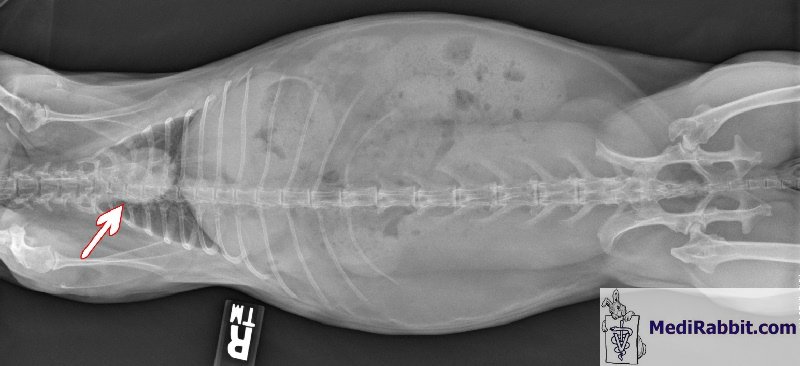
An X-ray of the abdominal region
of the rabbit is a commonly performed diagnostic examination, in case of
dyspnea (shortness of breath), a bad or persistent cough, a chest injury or
on suspicion of pneumonia. It will provide information about the shape and
the size of the heart and lungs. It can detect heart failure, emphysema, the
possible presence of pulmonary edema, the vascular pattern, the presence of
abscesses or neoplasia (e.g. thymoma, lung cancer), and other medical
conditions. This technique has its limitations though. Small malignant tumors
can be too small to be visible. Pulmonary embolism (blood clots to the lungs)
is not seen either, and require additional study.
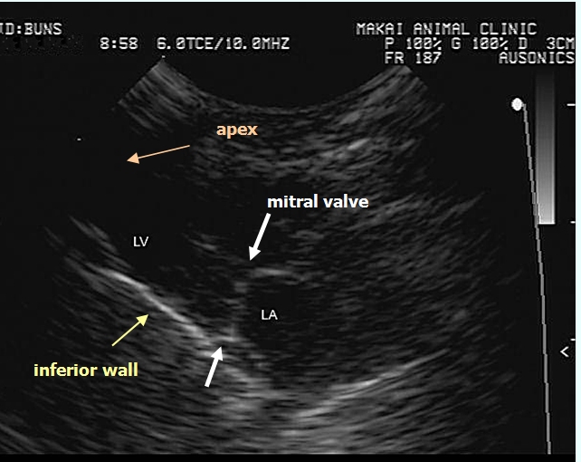
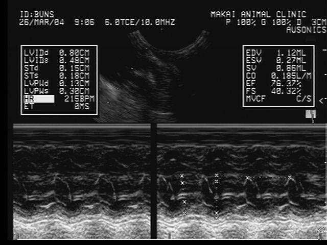
Rabbit ultrasound
examination or echocardiography
Most rabbits tolerate well the
harmless, non-invasive and widely available method of echocardiography, a
procedure that can be used without the use of sedative drugs, which can
modify the heart characteristics. The method is furthermore sensitive and
precise and the obtained images are of excellent quality. The rapid heartbeat
of rabbits and the small size of their hearts nevertheless require equipment
with a high frequency transducer (handheld recording probe) and a high frame
rate ultrasound machine. Echocardiography enables
detection of abnormalities in the heart structure (e.g. defective heart
valves, congenital defects), heart wall or chamber enlargement (e.g. heart
failure, cardiomyopathy), heart-wall motion, and allows the measurement of
the blood volume that is pumped from the heart with each beat. It can also
identify the accumulation of fluids in the pericardium (pericardial effusion)
or the presence of scar tissue throughout the pericardium. Special techniques, like M- or
TM-mode (M = movement, T = Time) ultrasound will provide information for the
analysis of wall and valve movements. The B-mode technique (B = brightness)
is used for examination of the anatomical relationships (e.g. the heart
structure, valves), while (color) Doppler ultrasonography will help determine
the direction of the blood flow and/or its velocity and can thus detect
turbulent flow due to narrowing or blockage of blood vessels.
Rabbit
electrocardiography (ECG or EKG)
Electrocardiography (ECG) is a
commonly used, non-invasive, simple and painless procedure that enables to
record electrical changes in the heart, by amplifying electrical impulses
that flow through the heart. Electrocardiography is used to evaluate and
manage causes of symptoms such as chest pain, dyspnea, palpitations,
arrhythmia, or syncope. The rhythm in a healthy rabbit
shows a sine. It excludes respiratory sinus arrhythmia (RSA), as there is no
influence of breathing on the flow of sympathetic and vagus impulses
to the sinoatrial node. The obtained electrocardiogram,
which shows a series of waves, will provide information about the pacemaker
(part that triggers each heartbeat), about the nerve conduction pathways of
the heart, and the rate and rhythm of the heart. The different waves are
called named P, Q, R, S, and T and follow in alphabetical order: • P
wave of the electrocardiogram is associated with the atrial contraction, • QRS
series of waves is associated with ventricular contraction, • P-Q
or P-R interval gives a value for the time taken for the electrical impulse
to travel from the auricle to the ventricle. • T
wave comes after the contraction. Electrocardiogram values for a
healthy rabbit:
Variation
is the values presented in the above table may indicate: • Abnormal P wave: right or left atrial
hypertrophy, atrial premature beat, hyperkalemia. • Abnormal QRS interval: right or left bundle
branch block, ventricular rhythm, hyperkalemia, among others. • Abnormal Q-T duration: hypocalcemia,
hypothyroidism, brain hemorrhages, congenital deformations, myocardial
infarction, myocarditis. • Abnormal T wave: hyperkalemia, hyperacute
myocardial infarction and left bundle branch block in case of a tall T wave;
ischemia, age, stress, pericarditis, intraventricular conduction delay,
electrolyte disturbance, in case of a small, flattened or inverted T wave. Rabbit cardiac disorders
Various disorders, including
congestive heart failure, cardiac myopathy (e.g. myocardial fibrosis), or
congenital heart disease (rare) like atrial or ventricular septal defects,
arrhythmia, valvular diseases, or vascular diseases have been observed in
rabbits. Acknowledgement Many thanks to
Tom Chlebecek, DVM, (Makai Animal Clinic, Kailua, HI), Frossie Economou, Kim
Chilson, and to Akira Yamanouchi, (Veterinary Exotic Information Network,
https://vein.ne.jp/), for giving their permission to use the pictures. Thank
you also Dr. Tom Chlebecek for his comments. Further
information M.V. Bray MV, WE. C. Weir EC, D. G. Brownstein, M.
L. Delano, (1992) Endometrial venous aneurysms in three New Zealand white
rabbits. Lab Anim Sci.; 42(4):360-2. Farkas,
A. J. Batey, S. J. Coker (2004) How to measure
electrocardiographic QT interval in the anaesthetized rabbit. J Pharmacol Toxicol Methods.;
50(3):175-85. L.C. St John, F. P. Bell (1990) Arterial fatty
acid-binding protein activity associated with dietarily-induced
and spontaneously occurring atherosclerosis in the rabbit (Oryctolagus
cuniculus). Comp Biochem Physiol
B.; 97(1):123-7. C. Kozma, W. Macklin, L.
M. Cummins, R. Mauer (1974) The anatomy, physiology
and biochemistry of the rabbit, in The Biology of the Laboratory Rabbit (Weisbroth et al., eds), pp
50-69. L. I. Kupferwasser, M.
R. Yeaman, S. M. Shapiro, C. C. Nast, A. S. Bayer
(2002) In vitro susceptibility to thrombin-induced platelet microbicidal protein is associated with reduced disease
progression and complication rates in experimental Staphylococcus aureus
endocarditis: microbiological, histopathologic, and
echocardiographic analyses. Circulation; 105(6):746-52. C. J. Orcutt (2000)
Cardiac and respiratory disease in rabbits. Proceedings of the British veterinary
Zoological Society (Autumn meeting) K. E. Quesenberry, J. W.
Carpenter, P. Quesenberry (2004) Ferrets, Rabbits
and Rodents: Clinical Medicine and Surgery Includes Sugar Gliders and
Hedgehogs, Elsevier Health, pp 211-216 R. S. Simons (1996) Lung morphology of cursorial and non-cursorial
mammals: lagomorphs as a case study for a pneumatic stabilization hypothesis.
J Morphol. 1996; 230(3):299-316. F. Harcourt-Brown, Textbook of Rabbit Medicine,
UK: Butterworth-Heinemann, 2001. |


e-mail: info@medirabbit.com